Wednesday, July 17, 2013

Esophageal disorders Congenital

2. DISORDERS OF THE OESOPHAGUS
 
2.1 CONGENITAL
 
ESOPHAGEAL ATRESIA
A  developmental defect is because of incomplete canalisation  of
esophagus. 
The defect is suspected on first feeding of  the  new
born.
 - There is choking coughing and cynosis on feeding.
 - There are five varieties of the observed defects as  shown  in
   Fig The variety C is the most common (85%).
 - The  diagnosis is  established by passage of  rubber  catheter
   through the nose and confirming the obstruction.
 
*  AP & Lat chest Xray will give the level of obstruction.
*  If Xray shows gas in GI Tract or Tracheo-Esophageal fistula is
   suspected .
 
TREATMENT
*   Pump suction of proximal pouch
*   Immediate assessment for other congenital defects
*   Surgical repair of the defect
 
Any questions be sent to drmmkapur@gmail.com
All older posts are stored in archives for access and review.
Visitors that follow may post contributions to the site.
To create consumer/provider engagement visit www.drmmkapur.blogspot.com

Wednesday, July 10, 2013

Esophagus 2 Physiology swallowing

The esophagus is a muscular tube fixed at its upper end to the cricoid cartilage and at its lower end to the diaphragm. This provides it transverse and longitudinal mobility, this mobility allows peristaltic wave to carry the bolus and this mobility also allows of intra thoracic organs to displace the esophogus without altering its food carrying functions.

The tube is lined by sqaumous epithilum with no keratin layer the submucosa contains elastic and fiberous tissue this forms the lamina propria the strongest and dependable layer for stitching in all surgery of esophagus. The lymph flow is not segmental and the submucouslymph plexus allows the lymph to flow long distance up and down, thus upper end cancers may show involvement of superior gastric glands and lower end carcinomas may show mediaitinal glands


1.1 PHYSIOLOGY 

The upper end of the oesophagus acts as a sphincter (UES) and  is
about 3 cm. in length and has a intraluminal resting pressure  of
20-60 mm of Hg.
 - With the act of swallowing this sphincter relaxes for 0.5 to 1
   second  as  the  bolus enters  the  oesophagus  the  sphincter
   contracts  and peristaltic waves of the oesophagus propel  the
   bolus to the lower sphincter (LES)
 - This (LES)  is the last 3 to 5 cm of the oesophagus that  acts
   as  a  sphincter though there is  no  demonstrable  anatomical
   structure like a sphincter.
 - This zone of the oesophagus develops a pressure of 10-20 mm of
   Hg  and  prevents regurgitation of stomach contents  into  the
   oesophagus.

Swallowing physiology
At the end of mastication the semisolid bolus is pushed by the postenor part of the tongue into the posterior oropharynx.
The soft palate (SP) rises to close of the opening of the neso pharynx. The hyoid and larynx move upward and the epiglottis classes of the larynx. These tow (SP+E) close of the nasal air passage and the larynx. The bolus is helped on its way towards the upper opening of the esophagus by the elevation of larynx.

The UES relaxes and the contractions of pharyngeal constrictors push the bolus into the esophagus. During the swallowing the breath is held. Further progress of the bolus is through the peristalsis of the esophagus.

Any questions be sent to drmmkapur@gmail.com
All older posts are stored in archives for access and review.
Visitors that follow may contributions to the site.
To create consumer/provider engagement visit www drmmkapur.blogspot.com
www.otmanage.blogspot.com 

Wednesday, July 3, 2013

Esophagus 1 Anatomy


OESOPHAGUS

1. ANATOMY
The oesophagus is a hollow muscular tube that begins at the lower
end  of the pharynx and ends in the stomach, it is 10 inches  (25
cm) in length.
 - Its maximum diameter in an adult is 2.5 cm and it  narrows  at
   its  junction with the pharynx (upper end) and stomach  (lower
   end).
 - It is  also narrow at the level of the 4th  thoracic  vertebra
   where the trachea bifurcates anterior to it .
 - The oesophagus is lined with mucosa (squamous cell) and has an
   inner circular muscular layer and an outer longitudinal layer.
 - In between these muscles, are the ganglion cells of Meissner's
   and  Auerbach plexus,the submucosa has the  Meissner's  neural
   plexus.
 - There is no serosa.

The  blood  supply  of  the  thoracic  oesophagus  is  from   the
oesophageal branches of the aorta, there are in addition branches
from  the  inferior  thyroid,  intercostal,  bronchial,  inferior
phrenic and left gastric arteries.

The   oesophagus  is  supplied  by  both  the   sympathetic   and
parasympathetic nerves.

The  lymphatics  arise  in the mucosa and form a  plexus  in  the
intermuscular  layer and the lymphatics drain to  the  lymphnodes
along the aorta

Any questions be sent to drmmkapur@gmail.com
All older posts are stored in archives for access and review.
Visitors that follow may post contributions to the site.
To create consumer/provider engagement visit www.drmmkapur.blogspot.com
www.otmanage.blogspot.om  

Wednesday, June 26, 2013

HEAD NECK 19 Short notes Epulis -Ludwig's angina


Chapter 14 page 1
Epulis
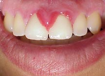
This  is an abnormal hyperplasia of the gums and is  produced  by
any  cause of chronic irritation (sharp edge of tooth cavity)  or
infection giant cell epulis is also because of inflammation.

Chapter 14 after 2.3

Torticollis
This  is the abnormal position of the head and neck caused  by  a
spasm  of  the  neck muscles. It can be  caused  by  an  abnormal
position while sleeping or an injury at birth. Treatment is  with
physiotherapy  and ultrasound. In the birth injury stretching  of
the muscles can also help.

Chapter 14 after 7.2

Adamantinoma
This is a multilocular cystic swelling usually seen in the  lower
jaw. It causes a bony expansion which can be seen on Xray. It  is
a slow growing tumour but invades tissue locally. The patient  is
20-30 years. Treatment is by local Excision.

Chapter 14 after 4.6

Ameloblastomar
This  tumour takes origen from the enamel organ of the teeth  and
can be seen in the upper or lower jaw also called admantinoma.  A
slow growing low-grade malignat tumour expands the jaw. X-ray  is
a  "soap  bubfle"  multiloculated cystic expansion  of  jaw.  The
lesion has to be excised and replaced with a bone graft.

Chapter 14 after 4.6

Odontoma
There are cysts that origen from ectodermal and mesodermal  teeth
producing tissue in the mouth. These cysts expend the jaw and are
usually benign.

Chapter 14 after 4.2

Ludwig's Angina
This  is  a severe cellulite of the submandibidor region  due  to
tooth  abscess  due to anaerobes and spirochetes the  ocdena  can
cause  respiratory obstruction. The patient needs  admission  and

 hugh dose of antibiotic and metronidazole.

Any questions be sent to drmmkapur@gmail.com
All older posts are stored in archives for access and review.
Visitors that follow may post contributions to the site.
To create consumer/provider engagement visit www.drmmkapur.blogspot.com  

Wednesday, June 19, 2013

HEAD NECK 18 malignant parotid

8.6.1 MALIGNANT MIXED TUMOURS

Malignant mixed tumours arise from a neoplastic transformation of
a  benign mixed tumour.  Patients are generally older,  malignant
lesions  are  usually  larger and any mixed  tumour  may  contain
within  it malignant elements and may result in  adeno-carcinomas,
squamous cell carcinoma or malignant spindle cell alternation.

With  the exception of mixed tumours; benign lesions of  salivary
tissues have no malignant potential.

CLINICAL PRESENTATION

Mixed  tumour may grow for years at a slow pace.   The  malignant
component  suddenly develops and there is a marked change in  its
course.

Fixation  of  glands to skin or underlying  structures,  palpable
nodes and involvement of the VIIth nerve are all diagnostic.

Excision of the mass and histologic examination are indicated  in

every case.

Any questions be sent to drmmkapur@gmail.com
All older posts are stored in archives for access and review.
Visitors that follow may post contributions to the site.
To create consumer /provider engagement visit www.drmmkapur.blogspot.com
www.otmanage.blogspot.com 

Monday, June 10, 2013

HEAD NECK 17 Parotid tumors

8.5 TUMOURS

8.5.1 MIXED TUMORS

A  common  lesion  of the salivary gland  is  the  mixed  tumours
(pleomorphic adenoma).

*  Eighty  percent of all benign tumours probably originate  from
   the myoepeithelial cells of the ducts.

*  Microscopic examination shows a network of strands of  spindle
   shaped and stellete cells.

*  In  one-third  of  cases loose  myxoid  pattern  predominates. 
   Half, in addition, have pseudocartilaginous structures.

Treatment  is  superficial  Parotidectomy  with  conservation  of
branches of the facial nerve.

8.5.2 PAILLARY CYSTADENOMA LYMPHOMATOSUM (WHARTHIN'S TUMOUR)

The lesion feels firm on examination and represent parotid tissue
sequestered in a lymphnodes.  It is the second most common benign
tumour of salivary tisse

The Histogenesis is unceertain


Treatment is surgical excision.

Any questions be sent to drmmkapur@gmail.com
All older posts are stored in archives for access and review.
Visitors that follow may post contributions to the site.
To create consumer/provider engagement visit www.drmmkapur.blogspot.com

Tuesday, June 4, 2013

HEAD NECK 16 Sialolithisis sialoadenitis

8.4 SIALOLITHIASIS (STONES)

Calculi  sometimes form within the ducts of salivary  glands  and
this occurs because of poor drainage and change of pH.

The symptoms are those of obstruction and distension of the gland
following  the  taking of food.  The gland may  enlarge  and  may
become palpable.

The  stone may be felt in the cheek in the parotid duct  and  the
floor of the mouth in the case of Submandibular duct symptoms are
of pain and swelling.

Treatment is by removal of the stone .

SIALOADENITIS

This occurs most often with invasion by bacteria.

Acute  parotitis occurs, with the symptoms of pain, swelling  and
fever progress is rapidly in these cases .

Obstruction  of the duct plays a causative role  dehydration  and
malnutrition also contribute.

Treatment  is  removal  of  causitive  factors  and  antibiotics. 
Drainage of abscess may be required.

Recurrent sialoadenitis also occur with repeated episodes of pain
and swelling with fever and its complication of orchitis has been

discussed elsewhere.

Any questions be sent to drmmkapur@gmail.com
All older posts are stored in archives for access and review.
Visitor that follow may post contributions to the site.
To create consumer/provider engagement visit www.drmmkapur.blogspot.com