

Chapter 14
page 1
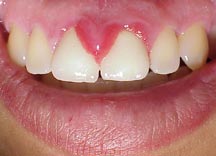
Epulis
This
is an abnormal hyperplasia of the gums and is produced
by
any
cause of chronic irritation (sharp edge of tooth cavity) or
infection giant cell epulis is also
because of inflammation.
Chapter 14
after 2.3
Torticollis
This
is the abnormal position of the head and neck caused by a
spasm
of the neck muscles. It can be caused
by an abnormal
position while sleeping or an injury at
birth. Treatment is with
physiotherapy and ultrasound. In the birth injury
stretching of
the muscles can also help.
Chapter 14
after 7.2
Adamantinoma
This is a multilocular cystic swelling
usually seen in the lower
jaw. It causes a bony expansion which
can be seen on Xray. It is
a slow growing tumour but invades tissue
locally. The patient is
20-30 years. Treatment is by local
Excision.
Chapter 14
after 4.6
Ameloblastomar
This
tumour takes origen from the enamel organ of the teeth and
can be seen in the upper or lower jaw
also called admantinoma. A
slow growing low-grade malignat tumour
expands the jaw. X-ray is
a
"soap bubfle" multiloculated cystic expansion of
jaw. The
lesion has to be excised and replaced
with a bone graft.
Chapter 14
after 4.6
Odontoma
There are cysts that origen from
ectodermal and mesodermal teeth
producing tissue in the mouth. These
cysts expend the jaw and are
usually benign.
Chapter 14
after 4.2
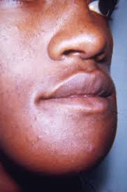
Ludwig's Angina
This
is a severe cellulite of the
submandibidor region due to
tooth
abscess due to anaerobes and
spirochetes the ocdena can
cause
respiratory obstruction. The patient needs admission
and
hugh dose of antibiotic and metronidazole.
Any questions be sent to drmmkapur@gmail.com
All older posts are stored in archives for access and review.
Visitors that follow may post contributions to the site.
To create consumer/provider engagement visit www.drmmkapur.blogspot.com


